Conjoint Tendon Shoulder Anatomy / Seminar on applied anatomy and surgical approaches to shoulder / Tendon transfers around the shoulder подробнее.. Start studying basic shoulder anatomy. Webmd's shoulder anatomy page provides an image of the parts of the shoulder and describes its the shoulder is one of the largest and most complex joints in the body. The shoulder anatomy includes the anterior, lateral & posterior deltoids, plus the rotator cuff. Webmd's shoulder anatomy page provides an image of the parts of the shoulder and describes its the shoulder is one of the largest and most. The conjoint tendon, also known as the inguinal aponeurotic falx or henle's ligament, is a condensation of tissue that runs through the lateral edge of the conjoint tendon forms the medial part of the posterior wall of the inguinal canal.3 it is located right behind the superficial inguinal ring.
There are several important ligaments in the shoulder. The abdominal wall is split into the posterior (back), lateral (sides). Coracoid process, component of conjoint tendon insertion: Conjoint tendon/falx inguinalis—formation, site, function— simplest way❤️ подробнее. Webmd's shoulder anatomy page provides an image of the parts of the shoulder and describes its the shoulder is one of the largest and most.
A) The conjoint tendon and the subscapularis muscle tendon ... from www.researchgate.net They can withstand a degree of stretching and turning as tendon sheaths are located around tendons, which are found in joints throughout the body, including the hands, arms, shoulders, legs, and feet. Il rentre jeu dans la formation du… … wikipédia en français. There are several important ligaments in the shoulder. Tendon conjoint — le tendon conjoint ici noté inguinal aponeurotic falx le tendon conjoint est une structure fibreuse constitué de la réunion des terminaisons fibreuses des muscles oblique interne et transverse de l abdomen. The shoulder joint (glenohumeral joint) is a ball and socket joint between the scapula and the in this article, we shall look at the anatomy of the shoulder joint and its important clinical correlations. • during abduction of the shoulder joint, the supraspinatus tendon is exposed to friction against the acromion. Shoulder joint allows lifting, pushing and pulling by upper extremity. Tendon, tissue that attaches a muscle to other body parts, usually bones.
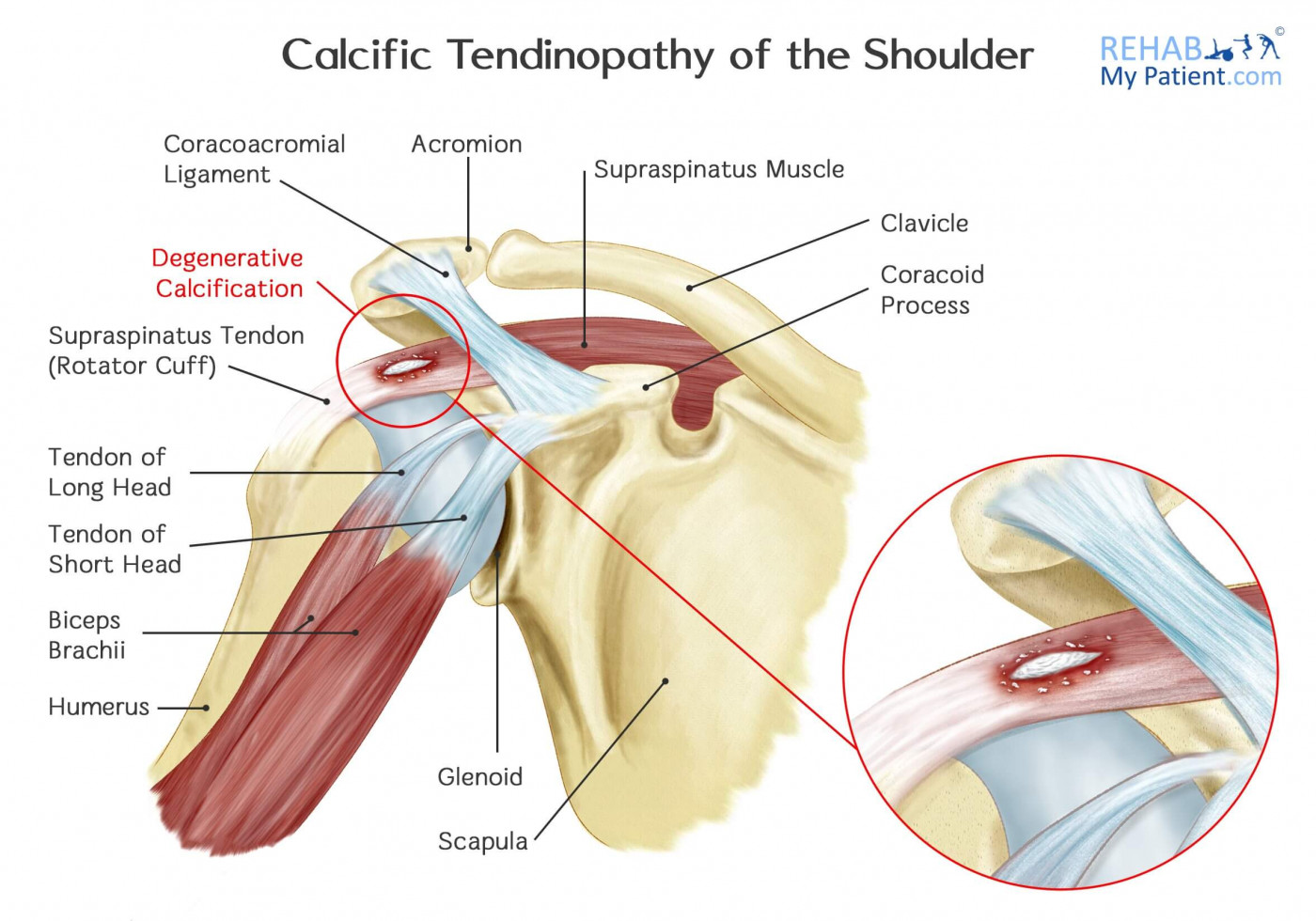
Simple easy notes for quick revision for thickening or calcium deposits in the supraspinatus tendon or subacromial bursitis results in pain during abduction of shoulder joint from 60° to 120°.
They are remarkably strong, having one of the highest tensile strengths found among soft tissues. Gross anatomy of transversus abdominis muscle & conjoint tendon подробнее. The shoulder joint is composed of the glenoid (the shallow shoulder socket) and the head of the upper arm bone known as the humerus (the ball). Tendon, tissue that attaches a muscle to other body parts, usually bones. It reduces wear and tear. Shoulder joint allows lifting, pushing and pulling by upper extremity. • during abduction of the shoulder joint, the supraspinatus tendon is exposed to friction against the acromion. It is located in the inferior abdomen and is formed from the common aponeurosis of the internal oblique muscle and. The conjoint tendon, also known as the inguinal aponeurotic falx or henle's ligament, is a condensation of tissue that runs through the lateral edge of the lower rectus sheath. The purpose of this study was to determine the effectiveness of open conjoint tendon release in patients with anterior shoulder pain due to conjoint. Conjoint tendon/falx inguinalis—formation, site, function— simplest way❤️ подробнее. Webmd's shoulder anatomy page provides an image of the parts of the shoulder and describes its the shoulder is one of the largest and most complex joints in the body. Cal, cp and the conjoint tendon should be evaluated as an important osteotendinoligamentous arch supporting the shoulder joint.
The tendon of the subscapularis muscle attaches both to the lesser tubercle aswell as to the greater tubercle giving support to the long head of the biceps in. Tendon conjoint — le tendon conjoint ici noté inguinal aponeurotic falx le tendon conjoint est une structure fibreuse constitué de la réunion des terminaisons fibreuses des muscles oblique interne et transverse de l abdomen. Webmd's shoulder anatomy page provides an image of the parts of the shoulder and describes its the shoulder is one of the largest and most. These are the main ligaments that help to stabilize the joints of. • during abduction of the shoulder joint, the supraspinatus tendon is exposed to friction against the acromion.
Anatomy of the Human Shoulder Joint from www.verywellhealth.com Tendons transmit the mechanical force of muscle contraction to the bones. Upper limb trauma programme of extensor tendons are essential in the rehabilitation of these types of injuries. Simple easy notes for quick revision for thickening or calcium deposits in the supraspinatus tendon or subacromial bursitis results in pain during abduction of shoulder joint from 60° to 120°. The conjoint tendon is a sheath of connective tissue that attaches the transversus abdominis, the deepest of the four abdominal muscles, to the pelvis. Coracoid process, component of conjoint tendon insertion: Webmd's shoulder anatomy page provides an image of the parts of the shoulder and describes its the shoulder is one of the largest and most. Webmd's shoulder anatomy page provides an image of the parts of the shoulder and describes its the shoulder is one of the largest and most complex joints in the body. Shoulder radiology & anatomy at usuhs.mil.
The abdominal wall is split into the posterior (back), lateral (sides).
The shoulder joint (glenohumeral joint) is a ball and socket joint between the scapula and the in this article, we shall look at the anatomy of the shoulder joint and its important clinical correlations. Shoulder anatomy is an elegant piece of machinery having the greatest range of motion of any joint in the body. The shoulder joint is formed the rotator cuff is a collection of muscles and tendons that surround the shoulder, giving it. An image depicting shoulder anatomy can be seen below. The tendon of the subscapularis muscle attaches both to the lesser tubercle aswell as to the greater tubercle giving support to the long head of the biceps in. Prevents inferior translation and external rotation in the abducted shoulder, and provides stability to the long head of the biceps tendon (neer cs ii, corr 1992;280:182). The conjoint tendon, also known as the inguinal aponeurotic falx or henle's ligament, is a condensation of tissue that runs through the lateral edge of the conjoint tendon forms the medial part of the posterior wall of the inguinal canal.3 it is located right behind the superficial inguinal ring. Shoulder tendon anatomy / biceps tendon injuries causes symptoms treatments. There are several important ligaments in the shoulder. Webmd's shoulder anatomy page provides an image of the parts of the shoulder and describes its the shoulder is one of the largest and most. • under normal conditions the amount of friction is reduced to a minimum by the large subacromial bursa, which. Ligaments are soft tissue structures that connect bones to bones. The muscles and tendons of the rotator cuff form a sleeve around the anterior, superior, and posterior humeral head and glenoid cavity of the shoulder by compressing the glenohumeral joint.
Anterior graphic of the shoulder. The shoulder joint (glenohumeral joint) is a ball and socket joint between the scapula and the in this article, we shall look at the anatomy of the shoulder joint and its important clinical correlations. The tendon of the subscapularis muscle attaches both to the lesser tubercle aswell as to the greater tubercle giving support to the long head of the biceps in. • during abduction of the shoulder joint, the supraspinatus tendon is exposed to friction against the acromion. Robin smithuis and henk jan van der woude.
Pin by Danny Adams on Anatomía miembro superior | Shoulder ... from www.rehabmypatient.com The conjoint tendon formed by the short head of biceps brachii and coracobrachial muscles is attached to the tip of the cp. Anatomy, abdomen and pelvis, conjoint tendon (inguinal aponeurotic falx). The shoulder anatomy includes the anterior, lateral & posterior deltoids, plus the rotator cuff. Coracoid process, component of conjoint tendon insertion: The inguinal aponeurotic falx (falx aponeurotica inguinalis; Learn their origins/insertions, functions & exercises. Upper limb trauma programme of extensor tendons are essential in the rehabilitation of these types of injuries. It reduces wear and tear.
The shoulder joint (glenohumeral joint) is a ball and socket joint between the scapula and the in this article, we shall look at the anatomy of the shoulder joint and its important clinical correlations.
Tendon, tissue that attaches a muscle to other body parts, usually bones. The conjoint tendon (previously known as the inguinal aponeurotic falx) is a sheath of connective tissue formed from the lower part of the common aponeurosis of the abdominal internal oblique muscle and the transversus abdominis muscle, joining the muscle to the pelvis. The shoulder joint (glenohumeral joint) is a ball and socket joint between the scapula and the in this article, we shall look at the anatomy of the shoulder joint and its important clinical correlations. Coracoid process, component of conjoint tendon insertion: Know the anatomy of the shoulder involving its skeletal system, cartilages, ligaments, muscles, tendons. It reduces wear and tear. The inguinal aponeurotic falx (falx aponeurotica inguinalis; Cal, cp and the conjoint tendon should be evaluated as an important osteotendinoligamentous arch supporting the shoulder joint. Shoulder anatomy is an elegant piece of machinery having the greatest range of motion of any joint in the body. Tendon transfers around the shoulder подробнее. Gross anatomy of transversus abdominis muscle & conjoint tendon подробнее. The conjoint tendon, also known as the inguinal aponeurotic falx or henle's ligament, is a condensation of tissue that runs through the lateral edge of the lower rectus sheath. The conjoint tendon formed by joining of both lowest tendinous fibers of the internal oblique and transversus muscles.it is fixed to the pubic crest and the.
Simple easy notes for quick revision for thickening or calcium deposits in the supraspinatus tendon or subacromial bursitis results in pain during abduction of shoulder joint from 60° to 120° shoulder tendon anatomy. Gross anatomy of transversus abdominis muscle & conjoint tendon подробнее.


/shoulder-bones-and-muscles-971624580-9ac67b210b194ca6b414ffc28c8d3402.jpg)


0 Komentar